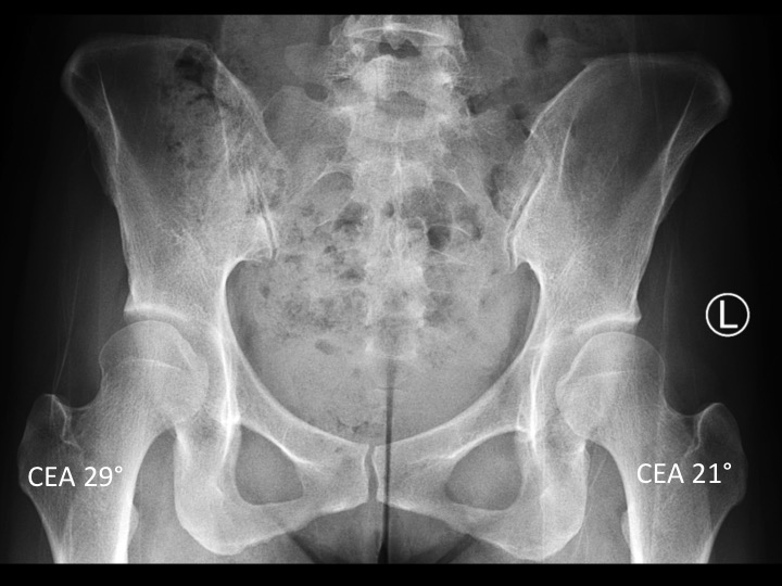
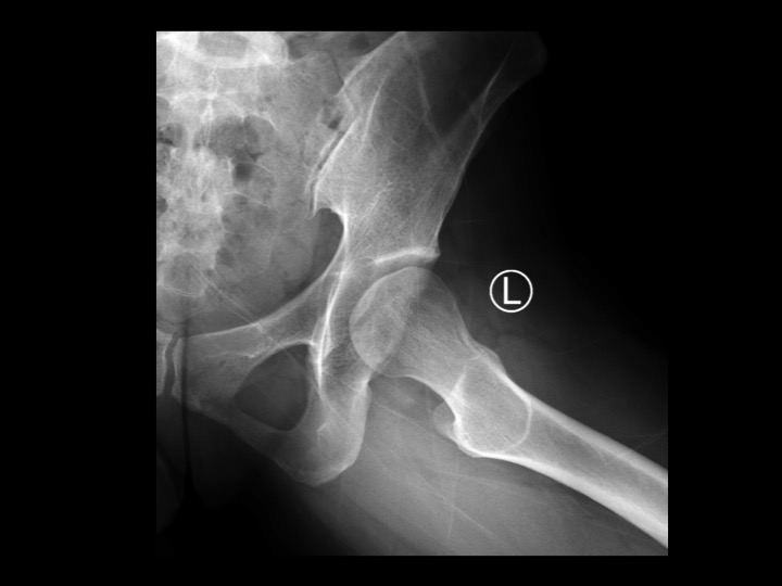
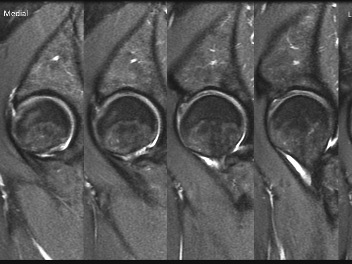
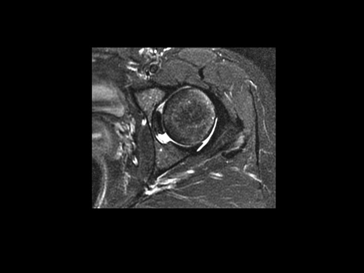
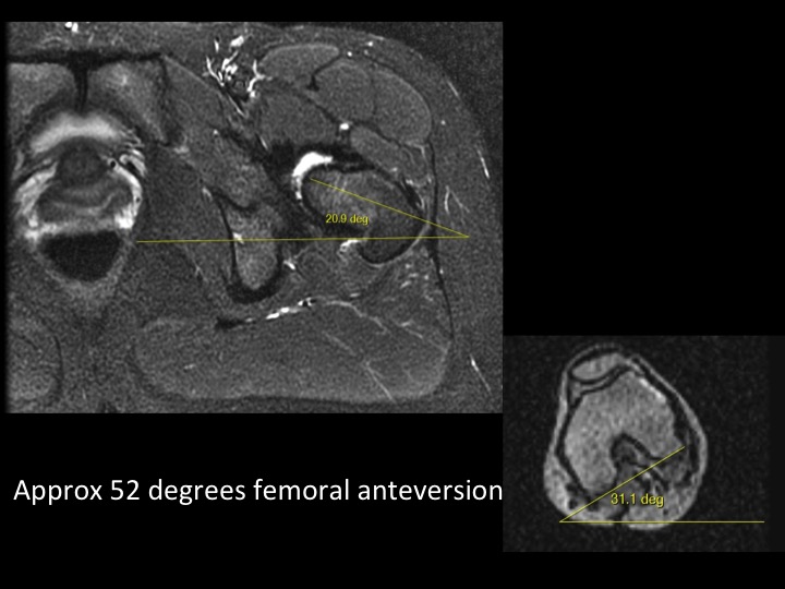
developmental dysplasia of the hip
Radiographs had been interpreted as normal, but show uncovering of the left > right femoral head as evidenced by the diminished left side center edge angle. The lateral XR shows typical loss of sphericity of the left femoral head. Sequential Sag MRI show a large delaminating tear of the left anterosuperior acetabular labrum; which is common in symptomatic DDH, though there is no secondary degenerative cartilage loss. Ax image redemonstrates the shallow acetabulum at the level of the center of the femoral head. Note the increased corrected femoral anteversion. [ Article ]
Rate Case : GoodGreatExcellentWOW !!!