Deprecated: Function eregi() is deprecated in C:\yeoman\xampp\htdocs\ocad\jview.php on line 24
Deprecated: Function eregi() is deprecated in C:\yeoman\xampp\htdocs\ocad\jview.php on line 24
Deprecated: Function eregi() is deprecated in C:\yeoman\xampp\htdocs\ocad\jview.php on line 26
Deprecated: Function eregi() is deprecated in C:\yeoman\xampp\htdocs\ocad\jview.php on line 28
Deprecated: Function eregi() is deprecated in C:\yeoman\xampp\htdocs\ocad\jview.php on line 29
Notice: Undefined variable: toRedirect in C:\yeoman\xampp\htdocs\ocad\jview.php on line 34 OCAD MSK (c) 2011
LOADING ...
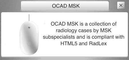
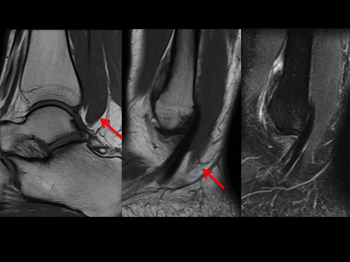
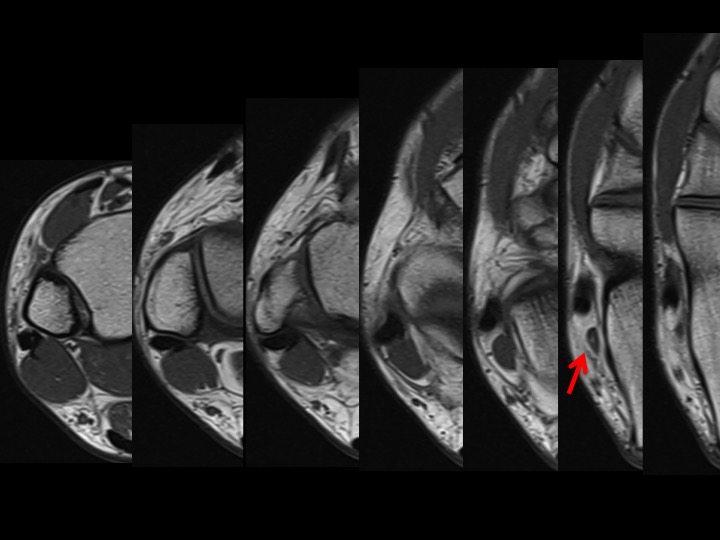
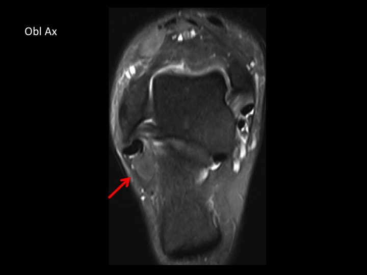
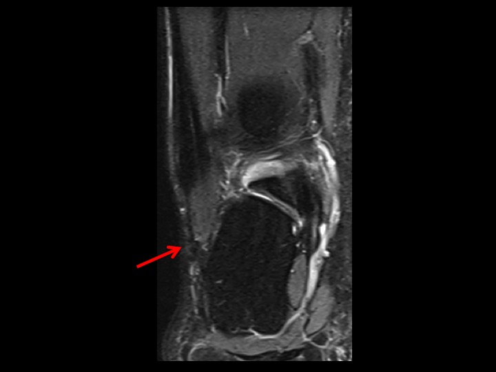
peroneus quartus Slide 1 shows a peroneal muscle (red arrow) which diverges from the course of the tendons at its caudal extent...meaning that it is an accessory peroneal muscle rather than a low lying peroneus brevis. Axial T1w images show this P Quartus to crowd the peroneal compartment posterior to the lateral malleolus, though it is only clear that it is a P Quartus (and not a low lying P brevis) as it veers off of the PB and PL tendons to insert onto the lateral calcaneus (this is the peronealcalcaneus variant of a P Quartus, which is the most common). These can be symptomatic or not. There is very minimal inframalleolar peroneal tenosynovitis. Though Obl Ax and Cor show the PQ pushing the tendons laterally within the confines of the retinaculum just caudal to the fibular tip, there is no tendinopathy. Her contralateral ankle was imaged, and though the PQ is often bilateral, in her case, this is unilateral. The PQ is also reported to be more frequent in men....so she is gloriously unique. [ Article ]