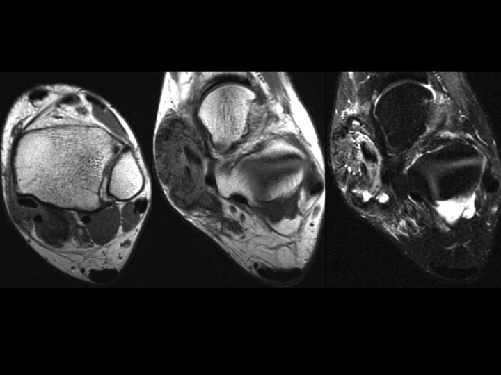
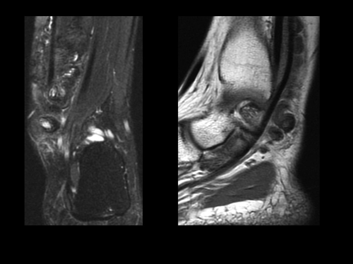
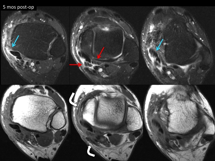
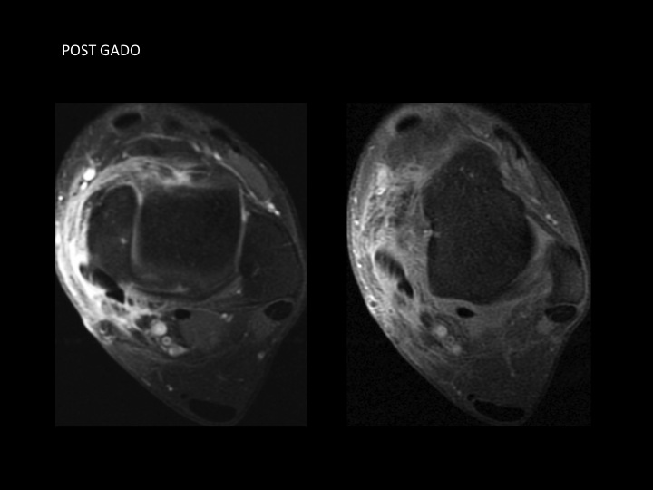
tenosynovial giant cell tumor
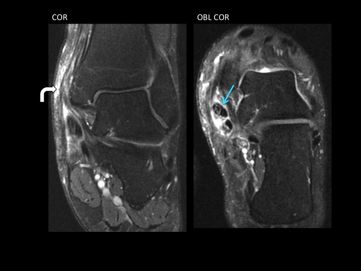
The pre-op images show the mass surrounds the PTT but extends around the FDL deep within the flexor compartment at the ankle and distal lower leg. Post-op there is thickening and retraction of the transected flexor retinaculum and stripping of the laciniate ligament (bent white arrows). There is residual TGCT in proximity to the FDL (red arrows), and there is new PTT tendinosis and partial tearing (blue arrows). The PTT is partially subluxed at the tibial margin, not surprising since the flexor retinaculum is gone; maybe this has contributed to the tendinosis. [ Article ]
Rate Case : GoodGreatExcellentWOW !!!