OCAD MSK

History

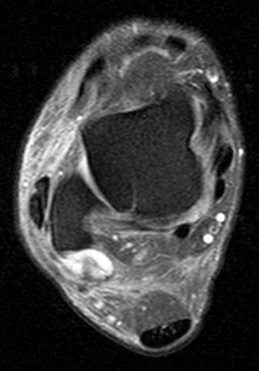
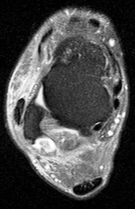
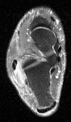
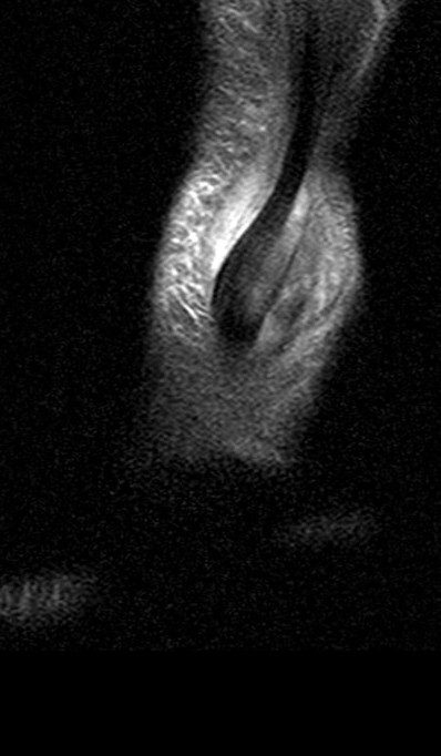
Pain. Follow up MRI. Old one showed hematoma.

Figure 1 for case peroneal dislocation
Figure 1
Figure 2 for case peroneal dislocation
Figure 2
Figure 3 for case peroneal dislocation
Figure 3
Figure 4 for case peroneal dislocation
Figure 4
Figure 5 for case peroneal dislocation
Figure 5

Discussion

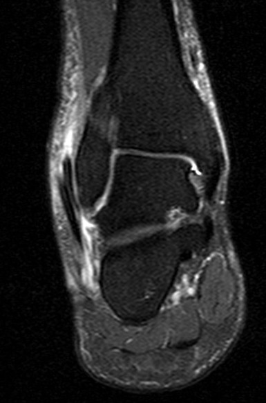
Both peroneals are dislocated around fibula. Retinaculum is torn. Initial case was missed. Patient walking around for nearly a year like this.

Diagnosis

peroneal dislocation

Phillip Tirman, MD
Courtesy