OCAD MSK
History
Arthroplasty with pretibial leg ulcer at the distal calf
Discussion
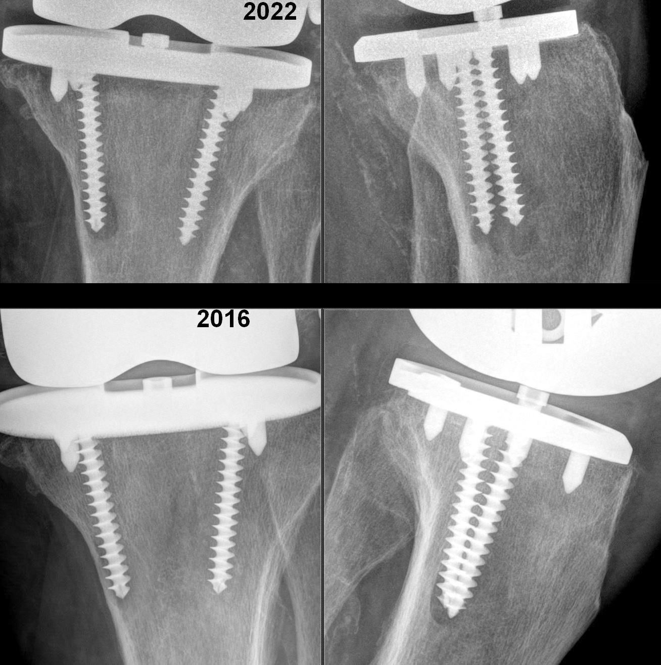
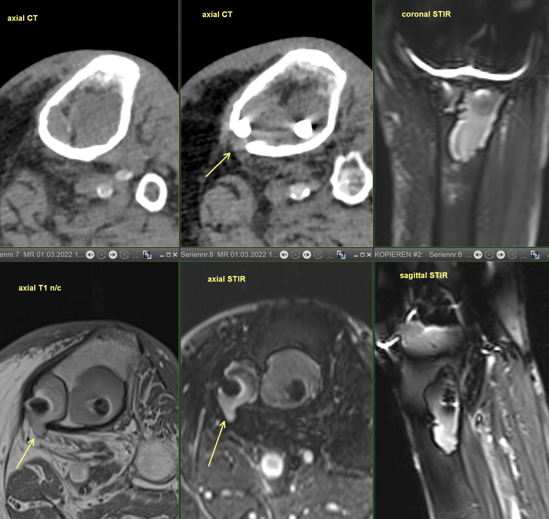
x-ray - Progressive osteopenia and lucency around tip of tibial screw. CT - lytic bone changes at the tibial prosthesis with a medial cortical defect. MR - cystic bone lesions at the tibial arthroplasty component extend out of the bone through a small defect. No surrounding marrow or soft tissue edema at this level.
Diagnosis
Presumed chronic material-related osteolysis