OCAD MSK
History
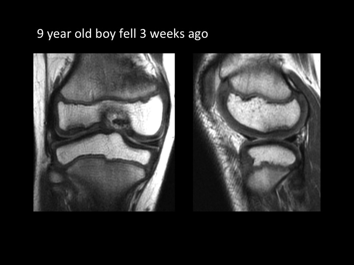
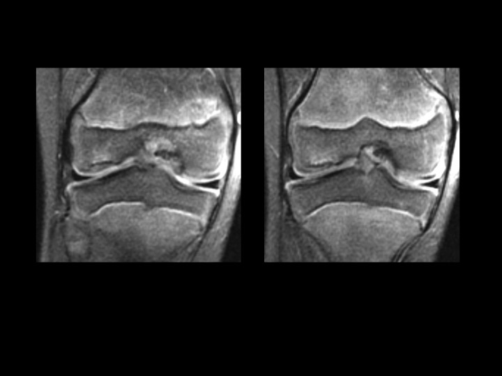
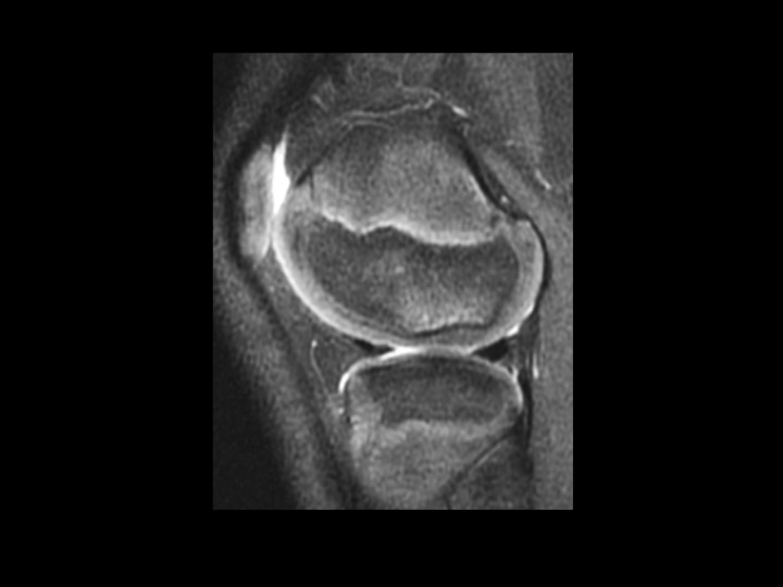
9M fell 3 weeks ago, evaluate osteochondral lesion
Discussion
The subchondral cortical flattening and minimal subchondral bright T2 signal with smooth, relatively thick overlying articular cartilage is not related to the injury 3 weeks prior. I considered possible chronic stable OCL, but I believe this looks like the normal variant ossification in the attached article. Any dissent? Reference article.
Diagnosis
normal developmental irregular ossification lateral femoral condyle