OCAD MSK
History
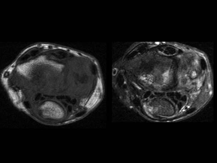
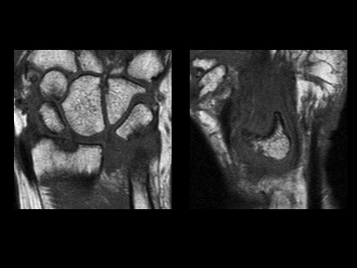
53F 6-8 mos pain with mass in carpal tunnel
Discussion
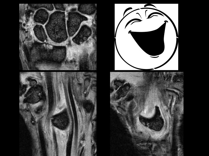
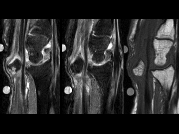
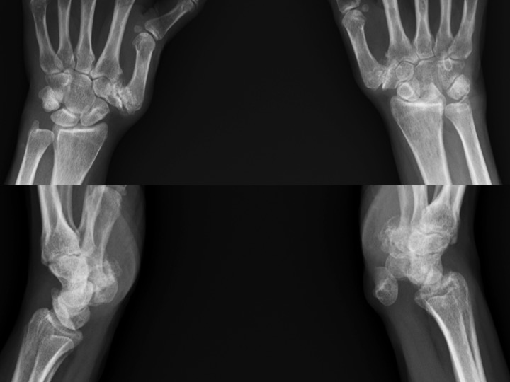
What makes this amusing (to me) is that the radiographs (slide 5) were done and reported (by me) 4 days prior to MRI. Patient claims prior work related injury, though this was not the focus of the office visit note. The hand surgeon interpreted the XR in his note as no scapholunate interval widening, no DISI (thats the funny part). MRI was ordered to evaluate mass in the carpal tunnel region. The patient moved a little because she was in alot of pain. The Sag PDFS images show the median nerve being stretched and compressed anterior to the chronically dislocated lunate. (the lunate seems to be happy) Reference article.
Diagnosis
Lunate dislocation