OCAD MSK
History
34F injury 1 week ago with pain and swelling
Discussion
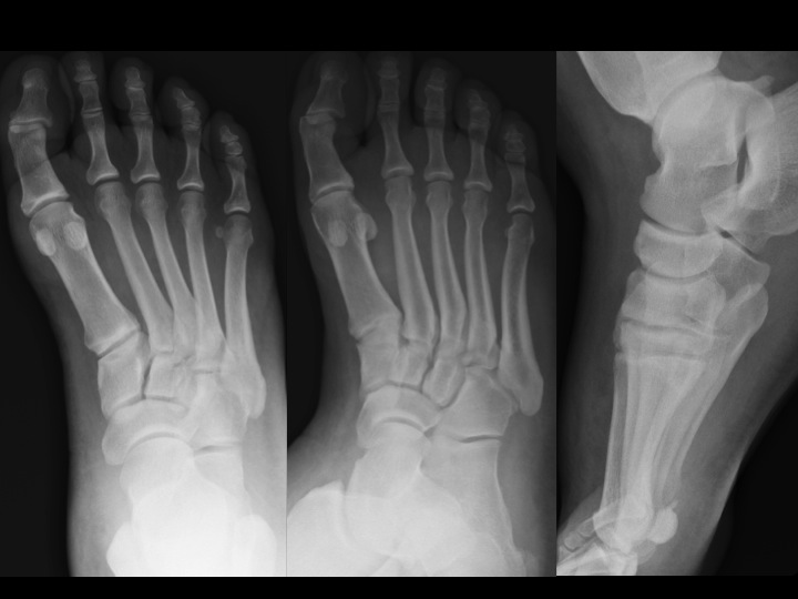
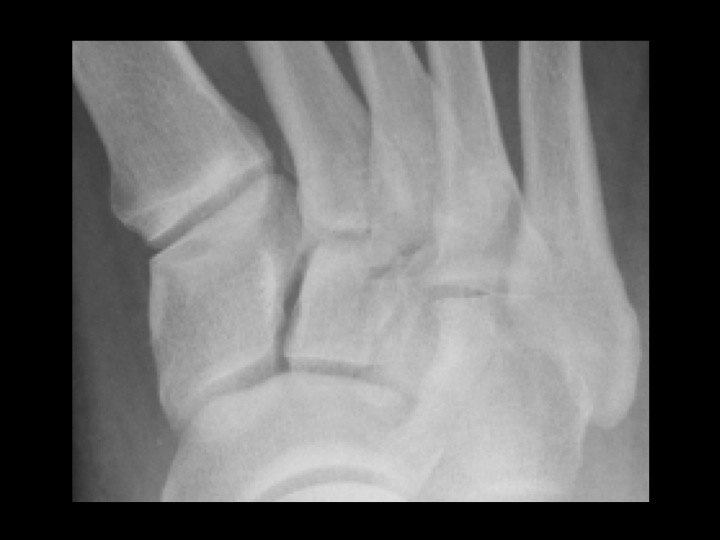
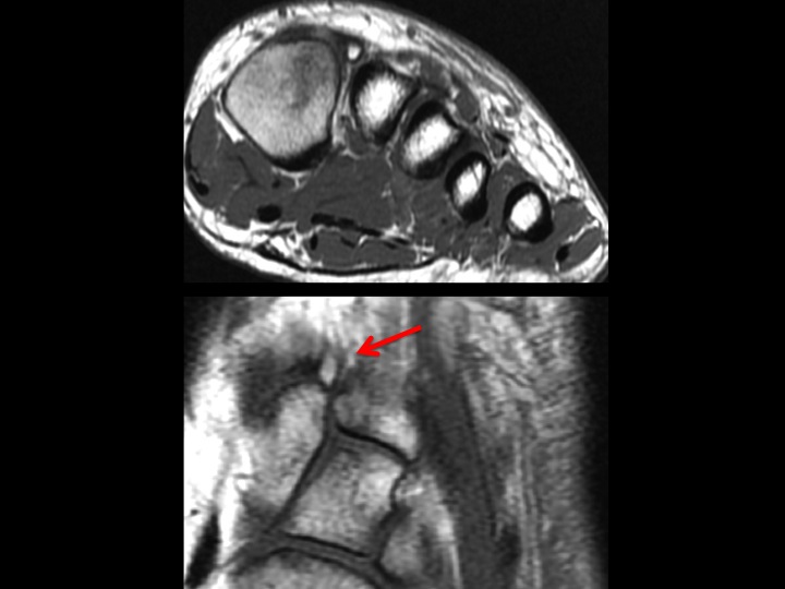
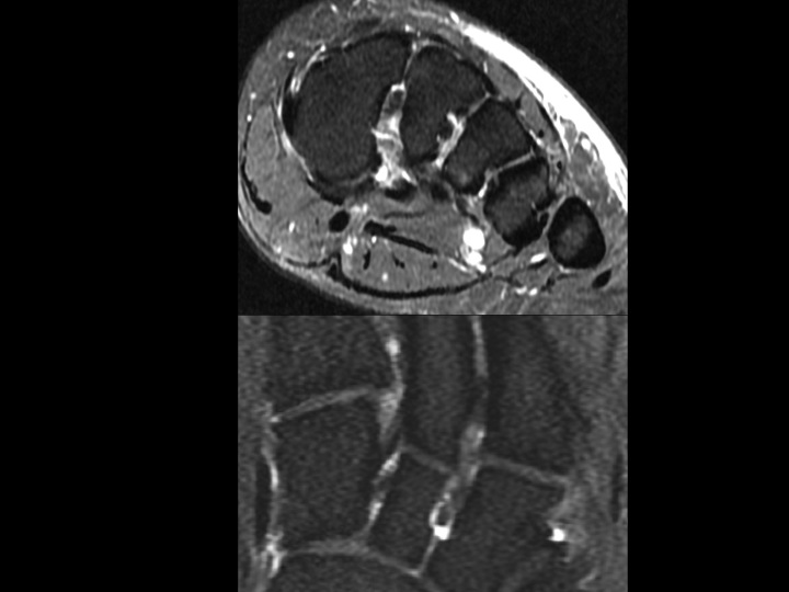
The tarsometatarsal alignment is preserved in the AP, Obl and Lateral radiographs, but there is marked dorsal foot swelling. These were non-weight-bearing. In the magnified AP view, there is a small fleck of bone in the proximal 1st intermetatarsal space suggesting a possible subtle Lisfranc injury. T1 weighted Cor and Ax images show the small, smoothly corticated os intermetatarseum, rather than a fracture fragment. The T2-fatsat images show the intact Lisfranc ligament complex. So what this patient has here is a boo boo.
Diagnosis
os intermetatarseum