OCAD MSK
History
Discussion
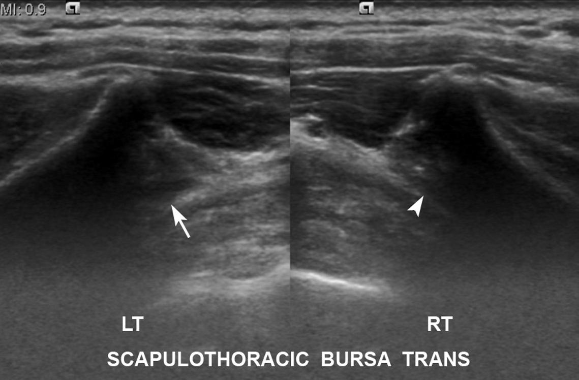
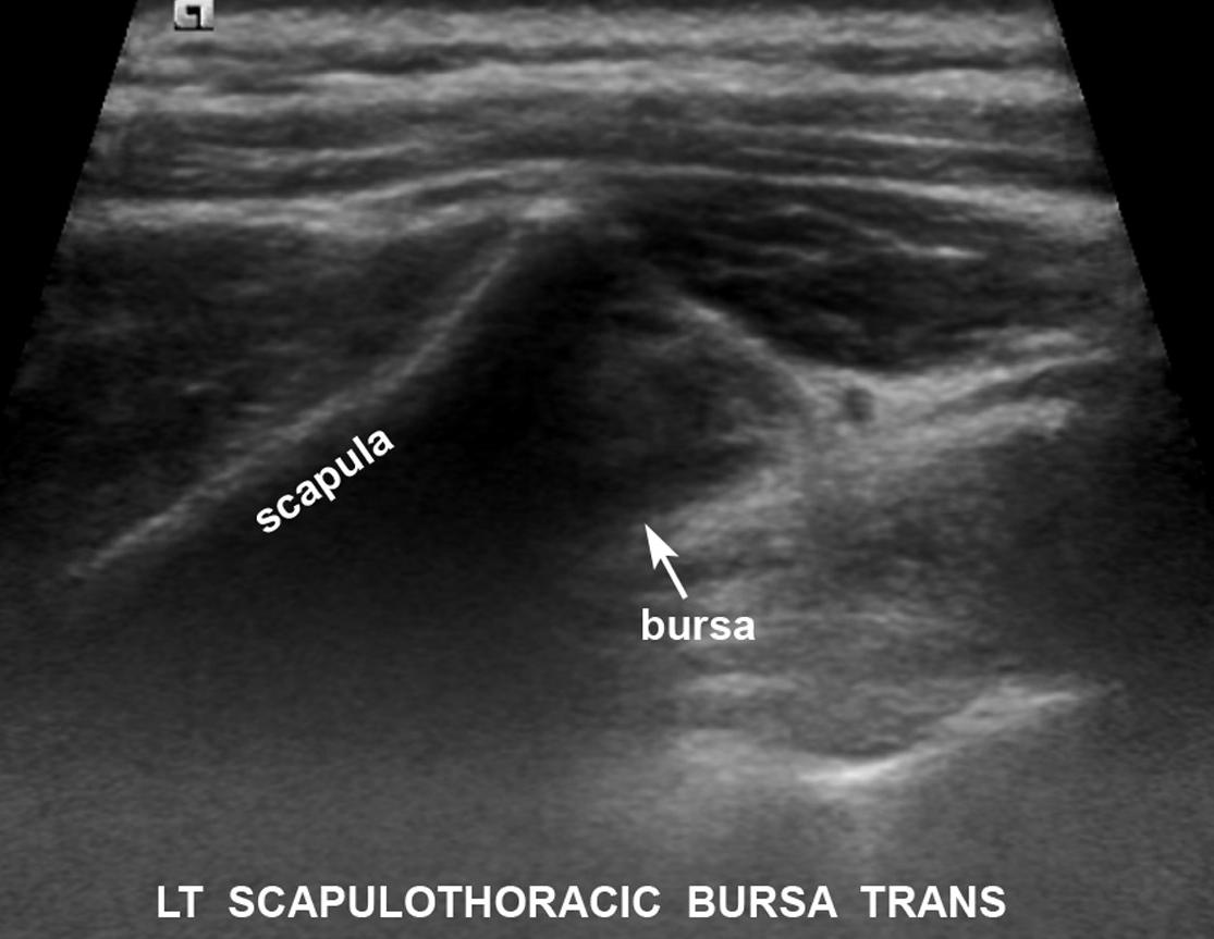
F25 with 4 months posterior pain left shoulder. Axial ultrasound images show hypoechoic thickening of the left scapulothoracic bursa (arrows) compared to normal right side (arrowhead). Note increased thickness of left scapulothoracic interval secondary to bursitis (accounting for clinically obvious swelling).
Diagnosis
Scapulothoracic bursitis